|

sicilymagazine.it
ha aggiunto
2 nuove foto.
19 maggio alle ore 22:30
·
NEW RECOMMENDATIONS ON HOW TO ASSESS PROSTHETIC HEART VALVES
by Guido Francesco Guida
Published on the forth of May in European Heart Journal –
Cardiovascular Imaging the first recommendations on multimodality
imaging assessment of prosthetic heart valves produced by the
European Association of Cardiovascular Imaging (EACVI), a registered
branch of the European Society of Cardiology (ESC).
Heart valve replacement is performed using mechanical or biological
prostheses. It is a device implanted in the heart of a patient with
valvular heart disease. When one of the four heart valves
malfunctions, the medical/surgical choice may be to replace the
natural valve with an artificial valve. This often requires
open-heart surgery. As opposed to valve replacement by open heart
nowadays can be performed
percutaneous aortic valve replacement (PAVR), also known as
transcatheter aortic valve implantation (TAVI) or transcatheter
aortic valve replacement (TAVR) that is the replacement of the
aortic valve of the heart through the blood vessels. The first
implantation of mechanical heart valve to human was performed by Dr.
Hufnagel on September 11, 1952 using the valve that he developed.
Heart valve replacement is performed using mechanical or biological
prostheses. It is estimated that by 2050, some 850 000 prosthetic
heart valves will be implanted every year in western countries.
Dysfunction of prosthetic heart valves is rare but can be life
threatening. When it does occur, it is crucial to determine the
cause as this will define what treatment is needed. The paper
provides the first recommendations on how to use multimodality
imaging to detect and diagnose prosthetic heart valve complications
and define treatment. When prosthetic heart valve complications are
suspected, the authors recommend:
First-line imaging with 2D transthoracic echocardiography (TTE)
2D and 3D TTE and transoesophageal echocardiography (TOE) for
complete evaluation
🔹
Cinefluoroscopy to evaluate disc mobility and valve ring structure;
🔹
Cardiac computed tomography (CT) to visualise calcification,
degeneration, pannus, thrombus;
🔹
Cardiac magnetic resonance imaging (CMR) to assess cardiac and
valvular function;
🔹
Nuclear imaging, especially when infective endocarditis is
suspected.
“In this paper we have underlined the incremental value of all
imaging modalities to evaluate prosthetic heart valves,” said
Professor Lancellotti. “Echocardiography should be used in the first
instance to detect any dysfunction. Non-echo imaging modalities can
be performed afterwards if more information is needed to establish
the cause and extent of complications.” Professor Lancellotti
concluded: “We have introduced new algorithms to help clinicians
diagnose and quantify prosthetic heart valve dysfunction. They are
easy to use and we hope will improve assessment and subsequent
management of patients so that when complications do occur, better
outcomes can be achieved.”
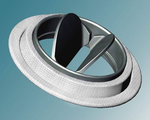
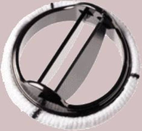
Picture source: numerik.math.tugraz.at
https://m.youtube.com/watch?v=ahkKZQBzss8
|
|